5 Neuroscience Research Instruments
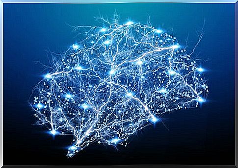
Neuroscience is a scientific discipline that studies the nervous system and how the different elements that make it up interact and give rise to behavior. It is a complex field of study that deals with neuronal functioning to behavior and therefore very broad. However, it is very useful to us when it comes to understanding how our behavior develops.
However, this discipline uses the scientific method to obtain knowledge through a series of research instruments in neuroscience. In fact, these are useful for both exploring the anatomy and functionality of the brain. Of course, each of them has certain advantages and disadvantages that make them suitable for certain situations and not for others.
For this reason, below we are going to briefly expose the instruments most used in neuroscience: the EEG, the MEG, the CT, the PET and the fMRI.
Electroencephalogram (EEG)
It is an instrument that measures how electricity flows through the cerebral cortex. When a neuron is activated, a passage of ions is produced through it that we can measure with a series of electrodes. These electrodes are placed directly on the scalp along with some type of substance that facilitates the passage of current. Thanks to this, we can capture neural activity in the form of waves.
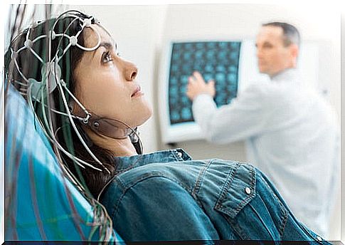
The EEG is one of the research instruments in neuroscience with great temporal capacity. However, its spatial ability is very poor. It is useful for us to relate wave patterns with certain processes, but if we want to locate them we must use another instrument.
An example of its use is during sleep phase investigations. This is because each of them corresponds to a specific wave pattern.
Magnetoencephalogram (MEG)
It is very similar to the EEG, but it does not capture the voltage changes, but the magnetic fields of the neurons. It is a physical principle that every electric current generates a magnetic field perpendicular to itself. Thanks to this, we can put receptors on the scalp that measure brain activity.
In addition, the structural anatomy of the cortex means that the magnetic field of some neurons does not leave the skull, while others do. This is useful to us to measure the activity of certain brain areas without noise or interference.
Compared to EEG, MEG has poorer temporal resolution. This is because the detection of the magnetic field has more delay. But it is true that it supposes a great improvement in spatial resolution, since we can know the location in which these magnetic fields have been generated.
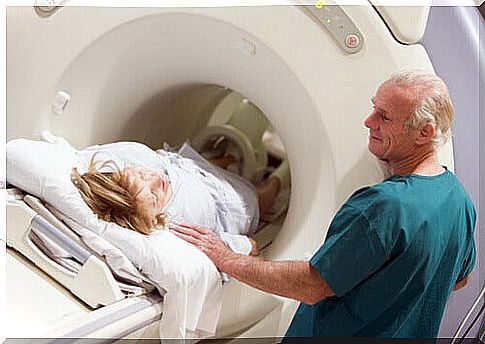
Computed Axial Tomography (CT)
It is one of the most useful neuroscience research tools for exploring the structural anatomy of the brain. It consists of passing a multitude of X-ray beams around the head from different angles. Once this is done, through a computer program, all the images are put together to have an image of the brain in 3D.
When crossing the human body, a certain part of the X-rays is absorbed by the structures that they cross. So, if we put a receiver on the other side, we can see a photograph of the X-ray residue. This will give us an image of the areas that it has crossed in a gray scale.
CT is a very useful technique to view brain anatomy and is very inexpensive, as well as being a simple practice. Still, it has certain drawbacks. The main and perhaps most serious is the invasiveness of the test. Some of the radiation is absorbed by the brain; this causes its use to be limited to avoid damage. In addition, today there are techniques with much better spatial and temporal resolution than CT, such as magnetic resonance imaging.
Positron Emission Tomography (PET)
PET allows you to determine the level of metabolic activity in each area of the brain. This is interesting for research, as it gives us great information about where brain activity occurs.
To accomplish this, the subject is injected with glucose bound to a radioactive marker (2-deoxy-D-glucose). This substance will travel to the brain, where the positrons of the radioactive isotope will react with electrons from surrounding atoms. Thus, they will destroy each other, releasing light in the process.
This light caused by the reaction of positrons can be captured by a receptor. In this way, it is possible to have an image of the areas where the brain has consumed more glucose.
This technique is usually used at the same time as a CT scan to know exactly the structures where glucose is being metabolized. PET has a high spatial resolution, but the temporal resolution leaves a lot to be desired, since you have to wait for the substance to be consumed by the brain. In general, this process occurs after the cognitive event that we want to measure.
In addition, it is one of the most invasive techniques within neuroscience research instruments. It involves the introduction of radiation directly to the brain, with the consequent danger to its structures. Therefore, it is only used in cases where it is very necessary.
Magnetic Resonance (MRI) and Functional Magnetic Resonance (fMRI)
Along with CT, MRI is one of the most widely used techniques in both neuroscience and medicine. MRI takes advantage of the physical fact that the atoms of certain substances in the human body react when an electromagnetic wave passes through them.
The MRI team uses a large magnet to orient the axis of all the hydrogen atoms in the brain in one direction. When the electromagnetic pulse ceases, all those atoms will reposition themselves, returning an energy signal that we can capture.
FMRI is a variant of the first one that allows us to measure brain activity and structure in real time, while the subject performs an activity with little time latency. Among the research instruments in neuroscience, it is possibly the one that gives us the best results both in space and time.
In addition, its invasiveness is totally null, since magnetic fields below a certain power do not damage the brain structure. However, its problem lies in its very high cost, both of the equipment and its maintenance. Getting an fMRI device costs around 5 million euros. Therefore, not all hospitals can afford to have one.
In this article, you have learned more about some of the neuroscience research instruments in use today. The study of this science is still in its early stages. However, thanks to these techniques, we know more and more about how the brain works.